


¿Por qué fallan las soluciones genéricas?
-
Problema con las soluciones genéricas
-
Errores comunes
¿Qué hacer?
-
Estrategias de apoyo general
-
Recomendaciones sobre estilo de vida
-
Recomendaciones nutricionales
-
Consideraciones sobre los suplementos
-
Nota sobre las nuevas pruebas/monitoreo
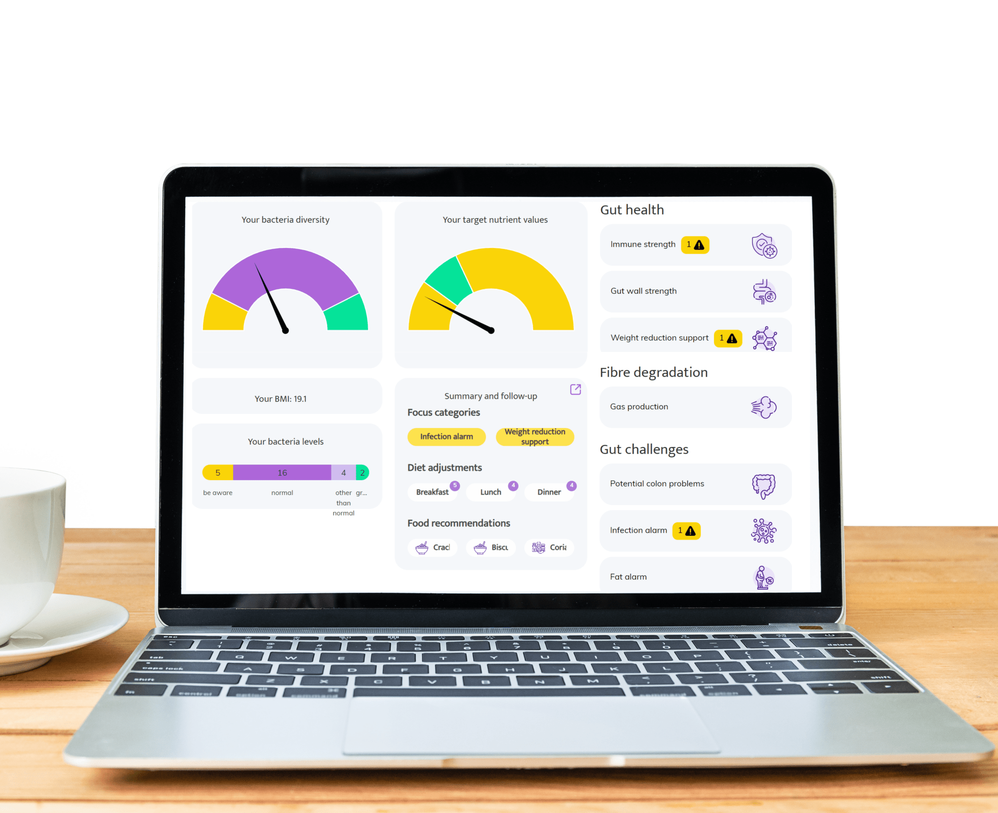
La importancia de las pruebas del microbioma intestinal
-
Por qué las pruebas son importantes
-
Cómo ayuda InnerBuddies
Evidencia médica
-
Nivel de evidencia científica
2 [débil; plausibilidad biológica emergente y evidencia humana mixta/insuficiente para la relevancia del riesgo de fibrosis, con datos causales y de utilidad clínica limitados]
-
Nota sobre la relevancia clínica
A continuación se presenta una lista de las publicaciones médicas más importantes relacionadas con esta condición específica.
| Title | Journal | Year | Link |
|---|---|---|---|
| The gut microbiome in pulmonary fibrosis | Frontiers in Immunology | 2022 | — |
| Microbiota and their metabolites in idiopathic pulmonary fibrosis | Nature Reviews Respiratory Medicine | 2021 | — |
| Intestinal dysbiosis and fibrosis: novel insights and potential mechanisms | Trends in Endocrinology & Metabolism | 2020 | — |
| Short-chain fatty acids regulate pro-fibrotic signaling in lung fibrosis | Science Translational Medicine | 2019 | — |
| Gut microbial metabolites modulate hepatic stellate cell activation and liver fibrosis | Hepatology | 2018 | — |
Preguntas frecuentes
¿Qué es MASLD y por qué es importante el riesgo de fibrosis?
¿Cómo puede el microbioma intestinal afectar el riesgo de fibrosis en MASLD?
¿Qué tienen que ver los SCFA y los ácidos biliares con el hígado?
¿Puede una prueba del microbioma predecir quién desarrollará fibrosis avanzada?
¿Qué mide la prueba InnerBuddies?
¿Están probados los biomarcadores del microbioma para el riesgo de fibrosis?
¿Cómo prepararse para una prueba de microbioma?
¿Cómo se realiza la prueba (tipo de muestra)?
¿Cuánto tiempo toma obtener los resultados?
¿Cómo deben interpretarse los resultados?
¿Qué puedo hacer para reducir el riesgo de fibrosis relacionado con el intestino?
¿Cómo puede la dieta afectar el eje intestino-hígado?
¿Debo empezar prebióticos o probióticos basándome en la prueba?
¿Con qué frecuencia debe repetirse la prueba del microbioma?
¡Escucha las opiniones de nuestros clientes satisfechos!
-
"Quiero contarles lo emocionada que estoy. Llevábamos unos dos meses con la dieta (mi marido come con nosotros). Nos sentíamos mejor, pero la verdadera mejoría se notó durante las vacaciones de Navidad, cuando recibimos un gran paquete navideño y nos saltamos la dieta durante un tiempo. Eso nos motivó de nuevo, ¡porque qué diferencia en los síntomas gastrointestinales y también en la energía que teníamos los dos!" - Manon, 29 años -
-
"¡¡¡Súper ayuda!!! Ya estaba bastante bien, pero ahora sé con certeza qué debo y qué no debo comer y beber. Llevo mucho tiempo luchando contra problemas de estómago e intestinos, espero poder deshacerme de ellos ahora." - Petra, 68 años -
-
"He leído su exhaustivo informe y sus consejos. Muchas gracias, me han resultado muy informativos. Presentados de esta manera, sin duda puedo seguir adelante. Por lo tanto, por ahora no tengo nuevas preguntas. Con mucho gusto tendré en cuenta sus sugerencias. Y le deseo mucha suerte con su importante labor." - Dirk, 73 años -
- Al seleccionar una opción, se actualiza toda la página.